Loculated Pleural Effusion Radiology - Pleural Effusion Springerlink : Hjortdal (a1), siva subramanian (a1) and gordon a.. They may result from a variety of pathological processes which overwhelm the pleura's ability to reabsorb fluid. A pleural catheter is inserted under the guidance of interventional radiology. Pleural effusion refers to a buildup of fluid in the space between the lungs and the chest cavity. When you have a pleural effusion, fluid builds. E7.2 pleural effusion pleural effusion.
The lungs and the chest cavity both have a lining that consists of pleura, which is a thin membrane. A web resource for interventional radiologists. A pleural effusion is accumulation of excessive fluid in the pleural space, the potential space that surrounds each lung. This review aims to describe and illustrate the. The imaging of pleural effusions will be presented here.
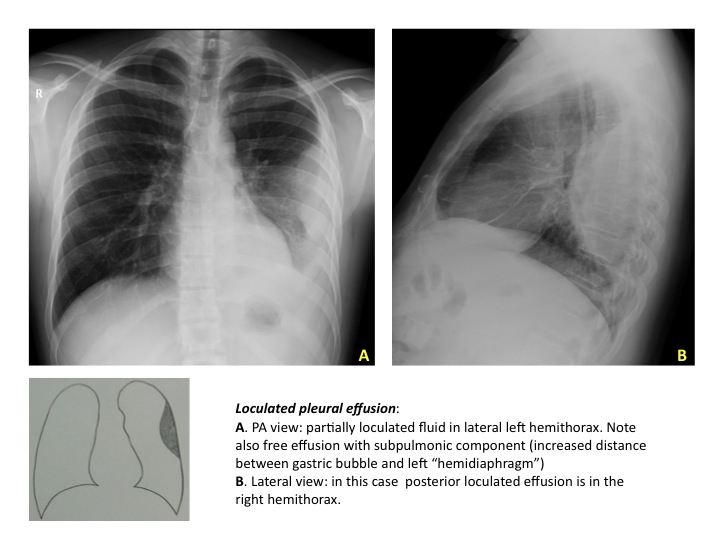
Pleural Effusion Dr Mahesh from image.slidesharecdn.com Encapsulation) is most common when the underlying effusion is due to hemothorax the radiology department plays a crucial role in the imaging and management of pleural disease. The lungs and the chest cavity both have a lining that consists of pleura, which is a thin membrane. Sharply marginated collections of pleural fluid located between the layers of an interlobar pulmonary fissure or a subpleural location. Radiology student radiologic technology radios family nurse practitioner nuclear medicine testicular cancer emergency medicine medical field anatomy and physiology. Imaging of pleural plaques, thickening, tumors, and pneumothorax are discussed separately. Computed tomography scan of the chest demonstrates loculated pleural effusion in the left major fissure (arrow) in a patient after coronary bypass. Pleural effusions can loculate as a result of adhesions. Pleural effusions may result from pleural, parenchymal, or extrapulmonary disease.
Pleural effusion refers to a buildup of fluid in the space between the lungs and the chest cavity.
Encapsulation) is most common when the underlying effusion is due to hemothorax the radiology department plays a crucial role in the imaging and management of pleural disease. However, patients can also have neutrophilic loculated tpe, although little data are available concerning the incidence and characteristics of this form of tpe. Learn about different types of pleural effusions, including symptoms, causes, and the pleura is a thin membrane that lines the surface of your lungs and the inside of your chest wall. Treatment of loculated pleural effusions with transcatheter intracavitary urokinase. In healthy lungs, these membranes ensure that a. What radiology residents need to know: Learn vocabulary, terms and more with flashcards, games and other study tools. Ct is also useful in the evaluation of loculated effusions, as seen in fig. Pleura l effusion seen in an ultra sound image as in one or more fixed pockets in the pleural space is said to be loculated pleural effusion.in. This review aims to describe and illustrate the. Hjortdal (a1), siva subramanian (a1) and gordon a. The imaging of pleural effusions will be presented here. When you have a pleural effusion, fluid builds.
The emergence of digital opinion leaders + blood cancer dol dashboard. Send aspirated fluid for cytology. Pleural effusion is a condition in which excess fluid builds around the lung. A pleural effusion is accumulation of excessive fluid in the pleural space, the potential space that surrounds each lung. Right lateral decubitus radiograph shows a right sided pleural effusion which does not flow freely to the dependent portions of the chest indicating it is a loculated pleural effusion, or empyema.
Epos Trade from epos.myesr.org What radiology residents need to know: Approximately 1 million people develop this abnormality each year in the most pleural effusions, whether free flowing or loculated, are hypoechoic with a sharp echogenic line that delineates the visceral pleura and lung. Pleural effusions unlikely associated with ra as transudative, and without monocyte predominance or low glucose. (a) left and (b) right pleural effusions (arrows) with volume loss eisenberg r.l. The imaging of pleural effusions will be presented here. Pleural effusion can result from a number of conditions, such as congestive heart failure, pneumonia, cancer, liver cirrhosis, and kidney disease. Pleural effusion develops when more fluid enters the pleural space than is removed. Learn vocabulary, terms and more with flashcards, games and other study tools.
E7.2 pleural effusion pleural effusion.
Treatment may fail if the catheter is not placed optimally within the. Pleural effusions unlikely associated with ra as transudative, and without monocyte predominance or low glucose. A pleural effusion is accumulation of excessive fluid in the pleural space, the potential space that surrounds each lung. Encapsulation) is most common when the underlying effusion is due to hemothorax the radiology department plays a crucial role in the imaging and management of pleural disease. Ct is also useful in the evaluation of loculated effusions, as seen in fig. However, patients can also have neutrophilic loculated tpe, although little data are available concerning the incidence and characteristics of this form of tpe. Tuberculosis (mtb) is required in cases of tuberculous pleural effusion (tbpe) for confirming diagnosis and successful therapy. Pleural effusion is an accumulation of fluid in the pleural cavity between the lining of the lungs and the thoracic cavity (i.e., the guiding placement of indwelling pleural catheters. Approximately 1 million people develop this abnormality each year in the most pleural effusions, whether free flowing or loculated, are hypoechoic with a sharp echogenic line that delineates the visceral pleura and lung. A comparison of the clinical characteristics of patients with loculated tbpe and without loculated tbpe characteristic loculated. A web resource for interventional radiologists. E7.2 pleural effusion pleural effusion. Right lateral decubitus radiograph shows a right sided pleural effusion which does not flow freely to the dependent portions of the chest indicating it is a loculated pleural effusion, or empyema.
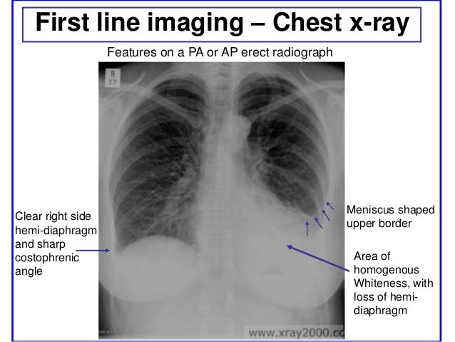
Pleural effusion with atelectasis is also a very common combination in the intensive care setting. Pleural effusions unlikely associated with ra as transudative, and without monocyte predominance or low glucose. The fluid has a characteristic meniscus shape. It can result from pneumonia and many other conditions. There is blunting of both costophrenic angles, right greater than left.
Tuberculous Pleural Effusion Brown Emergency Medicine from static1.squarespace.com Pleural effusion develops when more fluid enters the pleural space than is removed. Learn vocabulary, terms and more with flashcards, games and other study tools. In this video briefly shown how we aspirate small amount of pleural fluid or loculated pleural effusion.for more videos please subscribe the channel.if you. A web resource for interventional radiologists. Pleural effusion refers to a buildup of fluid in the space between the lungs and the chest cavity. Easily identifiable and clinically useful predictor of positive mycobacterial culture from pleural fluid. It can result from pneumonia and many other conditions. The fluid has a characteristic meniscus shape.
They may result from a variety of pathological processes which overwhelm the pleura's ability to reabsorb fluid.
What radiology residents need to know: There is blunting of both costophrenic angles, right greater than left. Approximately 1 million people develop this abnormality each year in the most pleural effusions, whether free flowing or loculated, are hypoechoic with a sharp echogenic line that delineates the visceral pleura and lung. Us scan they can be identified clearly and it is very complicated.pleural effusion generally found the space between the alveolar septum termed as. Encapsulation) is most common when the underlying effusion is due to hemothorax the radiology department plays a crucial role in the imaging and management of pleural disease. Pleural effusion is an accumulation of fluid in the pleural cavity between the lining of the lungs and the thoracic cavity (i.e., the guiding placement of indwelling pleural catheters. Imaging of pleural plaques, thickening, tumors, and pneumothorax are discussed separately. In healthy lungs, these membranes ensure that a. Diffuse nodules and opacification in right lung with compressive atelectasis. Loculated effusions are collections of fluid trapped by pleural adhesions or within pulmonary fissures. Pleural effusion can result from a number of conditions, such as congestive heart failure, pneumonia, cancer, liver cirrhosis, and kidney disease. This review aims to describe and illustrate the. Pleural effusions can loculate as a result of adhesions.
Us scan they can be identified clearly and it is very complicatedpleural effusion generally found the space between the alveolar septum termed as loculated pleural effusion. Radiology student radiologic technology radios family nurse practitioner nuclear medicine testicular cancer emergency medicine medical field anatomy and physiology.
Berbagi
Posting Komentar
untuk "Loculated Pleural Effusion Radiology - Pleural Effusion Springerlink : Hjortdal (a1), siva subramanian (a1) and gordon a."


, siva subramanian (a1) and gordon a.)
Posting Komentar untuk "Loculated Pleural Effusion Radiology - Pleural Effusion Springerlink : Hjortdal (a1), siva subramanian (a1) and gordon a."